
CLINICAL PRACTICE REAPPRAISAL
NAVIGATING THE ELECTROPHYSIOLOGICAL LANDSCAPE OF CORTICAL MYOCLONUS
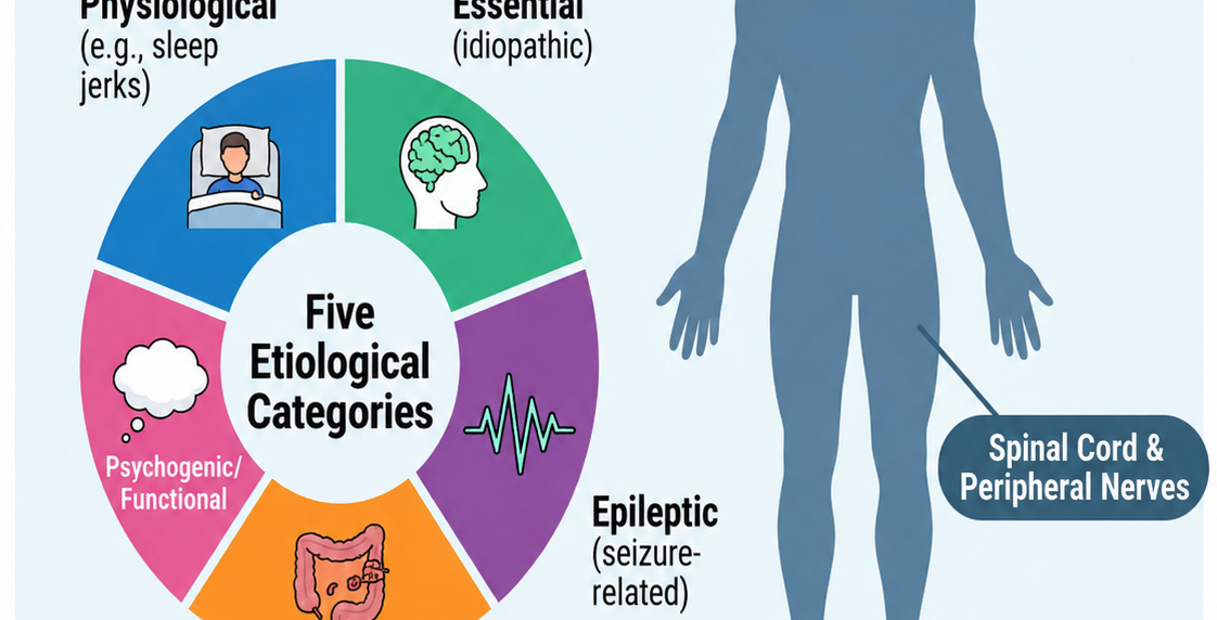
1. The pathophysiological basis and clinical urgency of cortical myoclonus
Cortical myoclonus (CM) must be reappraised not merely as a symptom of sensorimotor cortex hyperexcitability, but as a failure of complex inhibitory networks. While traditionally attributed to abnormal neuronal discharges within the neocortex, modern neurophysiology identifies a more nuanced "thalamo-cortical volley." This involves the primary somatosensory cortex (S1, specifically area 3b for cutaneous input and area 3a for proprioceptive afferents) and the primary motor cortex (M1). Critically, recent evidence suggests that cerebellar disinhibition (a reduction in the inhibitory output to the thalamus) may be a primary driver of this cortical state. Distinguishing CM from subcortical or peripheral counterparts is strategically vital; it dictates whether a clinician targets GABAergic dysfunction or structural spinal lesions, and it provides a prognostic window into the severity of neurodegenerative or post-hypoxic encephalopathies.
Anatomical scaffolding: The CM generator is rarely an isolated focal point, but often represents a pathological oscillation within a network. The following table synthesises the anatomical generators across the neurophysiological spectrum:
The diagnostic challenge
Newly onset myoclonus remains a profound challenge. The "stepwise approach" currently utilised often fails because the clinical semiology of CM can overlap significantly with subcortical variants. Furthermore, the reliance on a single positive test is a trap; many "gold standard" criteria are fulfilled only by a minority of patients. A true clinical reappraisal necessitates moving toward a multimodal evidence model where electrophysiological tools are viewed as pieces of a larger pathophysiological puzzle rather than binary "yes/no" indicators.
2. Deconstructing the "gold standard" criteria: a critical reappraisal
We must acknowledge a hard truth in movement disorder speciality: the evidence base for "gold standard" CM criteria is far less robust than traditionally assumed. Clinicians often find that these markers are absent even in confirmed cases, leading to frequent false-negatives.
- Giant somatosensory evoked potentials (SEPs): Standard definitions of "giant" SEPs typically cite a P25 amplitude >8.6 μV or an N33 amplitude > 8.4 μV. However, these thresholds are subject to clinical judgment regarding skull thickness and brain state.
- The "so what?" layer: S1 hyperexcitability (the giant SEP) does not always "drive" M1 motor output. This explains why SEPs can be giant in non-myoclonic conditions such as pain, multiple sclerosis, and motor neuron disease. Conversely, in corticobasal syndrome (CBS), SEPs are frequently normal or show loss of morphology despite clear cortical jerks, suggesting a distinct cortical-subcortical loop involvement where the motor cortex is reached via alternative, non-thalamo-cortical pathways.
- Jerk-locked back-averaging (JLBA): JLBA remains a cornerstone for identifying a pre-myoclonic EEG transient, but its technical pitfalls are numerous.
- Latency variations: While a ~20 ms latency (EEG spike to EMG burst) is characteristic of fast corticospinal transmission in PMEs, significantly longer latencies are observed in coeliac disease, Alzheimer’s, and Creutzfeldt–Jakob disease (CJD), where the drive may utilise slower descending pathways.
- The far-field pitfall: Positive-deflection EEG transients in JLBA may actually represent far-field potentials (P14) generated subcortically, potentially leading to a misdiagnosis of a cortical generator.
- The C-reflex (long-latency reflex): The pathological C-reflex must be differentiated from physiological long-latency reflexes (LLRs). In healthy subjects, LLR-I (35–46 ms) is seen only during contraction; in CM, the C-reflex (39–55.3 ms) may appear even at rest.
Best Practices: C-reflex and SEP protocol
- Stimulation: 1 Hz stimulation is mandatory for SEPs to avoid the extinction of enlarged middle and late components associated with higher frequencies.
- Intensity: Median nerve stimulus at 10% above motor threshold.
- Conditioning: Test the C-reflex both at rest and during active motor activation to distinguish it from the physiological LLR-II (45–58 ms).
3. Advanced markers and ancillary electromyographic features
The evolution from simple polygraphy to complex signal processing allows for the identification of "secondary" markers that bridge the diagnostic gap when JLBA is negative.
- High-frequency oscillations (HFOs): An emerging and specific biomarker for benign adult familial myoclonus epilepsy (BAFME) is the presence of HFOs (>400 Hz) within the giant SEP. These oscillations likely reflect abnormal thalamo-cortical circuit activity and provide high specificity for this phenotype compared to other PMEs.
- Cortico-muscular coherence (CMC) and phase-lag analysis: CMC identifies connectivity, but we must exercise caution: it does not inherently prove drive.
- The "so what?" layer: To confirm that M1 is driving the muscle, clinicians must employ phase-lag analysis. Without this, CMC only demonstrates functional linkage, which may be afferent (muscle to cortex) rather than efferent. Notably, Unverricht–Lundborg disease is associated with alpha-band coherence, while sialidosis typically exhibits gamma-band drive.
TMS and GABAergic dysfunction: Transcranial magnetic stimulation (TMS) provides the underlying "why" for CM. A consistent finding across CM aetiologies is the impairment of short-interval intracortical inhibition (SICI). This reduction in SICI directly correlates with GABA-A-mediated disinhibition, offering a physiological explanation for the hyperexcitable state that unites various genetic and symptomatic disorders.

4. Aetiology-specific neurophysiological profiles
The neurophysiological "signature" shifts as a function of the underlying disease state. Recognising these patterns allows for earlier syndromic classification.
- Progressive myoclonus epilepsies (PME): Characterised by photosensitivity, poly-spikes-and-waves, and nearly universal giant SEPs. In Lafora Disease, occipital spikes may be prominent.
- Post-hypoxic myoclonus: In the acute stage, myoclonic status epilepticus (MSE) presents with burst suppression. In the chronic Lance–Adams Syndrome (LAS), the myoclonus is often a mix of cortical and reticular (brainstem) reflex origin.
- Creutzfeldt–Jakob Disease (CJD): Shows diffuse periodic triphasic waves on EEG. JLBA often reveals extremely long latencies (tens of ms), suggesting a cortical-subcortical loop rather than a pure corticospinal drive.
Red flags (atypical pathologies):
- Uraemia: Induces reticular, stimulus-sensitive myoclonus due to medullary toxicity.
- Lennox-Gastaut Syndrome: Often demonstrates longer JLBA latencies, similar to neurodegenerative variants.
- Drug toxicity: ASMs at high doses can paradoxically worsen CM.
5. An integrated framework for increasing diagnostic reliability
A transition from "single-test reliance" to a "multimodal evidence" model is non-negotiable for the modern specialist.
Personalised polygraphy: Standardised muscle sets are often insufficient. The clinician must tailor the recording to the patient’s clinical distribution, as focal distal jerks require different montages than generalised axial involvement, to accurately capture the generator.
Clinical pearls for the specialist
- Identify the drive: Do not rely on CMC alone; use phase-lag analysis to ensure you are seeing efferent cortical drive.
- Scepticism of recruitment: Do not assume "cranial-to-caudal" progression confirms a cortical origin; the evidence for this is currently clear only in a minority of cases.
- Stimulus frequency: Always use 1 Hz for SEPs to capture the late components that signify the full thalamo-cortical volley.
Final synthesis
The current neurophysiological framework for CM is evolving from a set of rigid criteria into a nuanced study of network disinhibition. While JLBA and giant SEPs remain useful, they are frequently absent. Diagnostic reliability is only achieved by synthesising clinical semiology with a totality of data, including HFOs, SICI impairment, and directional coherence measures. Larger, standardised studies are imperative to establish the true sensitivity of these markers, but in practice, the integration of multiple tests remains our most powerful tool for navigating the hyperexcitable cortex.